Dural Arteriovenous Fistula Referral and Research Portal
Sree Chitra Tirunal Institute for Medical Sciences and Technology, Trivandrum
An Institution of National Importance, Department of Science and Technology, Govt. of India
Image Gallery
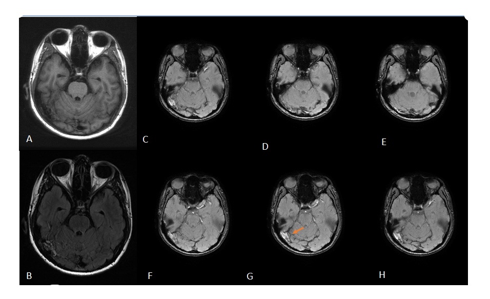
T1 appears normal(A) , FLAIR axial images hyperintensity in the region of right transverse sinus(B) , C-H shows susceptibility weighted MRI images that reveal hyperintense vascular structures (arrow) in the region of right transverse sinus, suggesting dural fistula
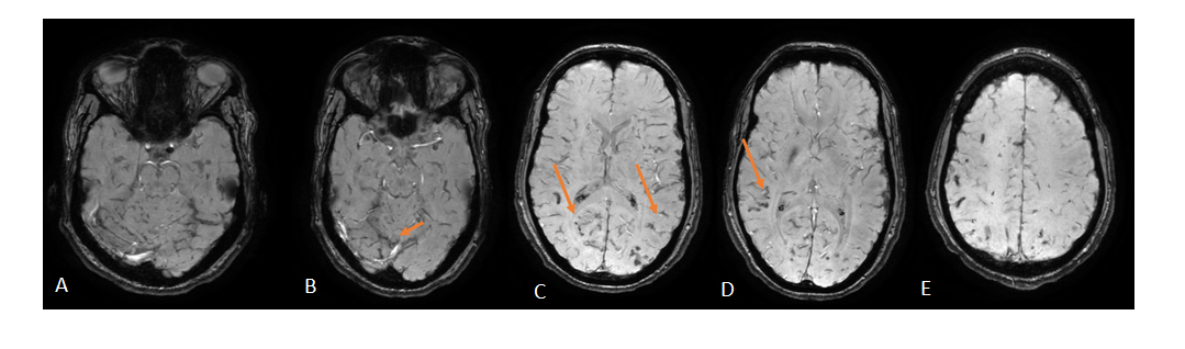
The reflux into straight sinus is evident suggesting aggressive dural fistula. Signs of venous congestion in the form of dilated venous structures are seen in the bilateral cerebral hemispheres(arrow)
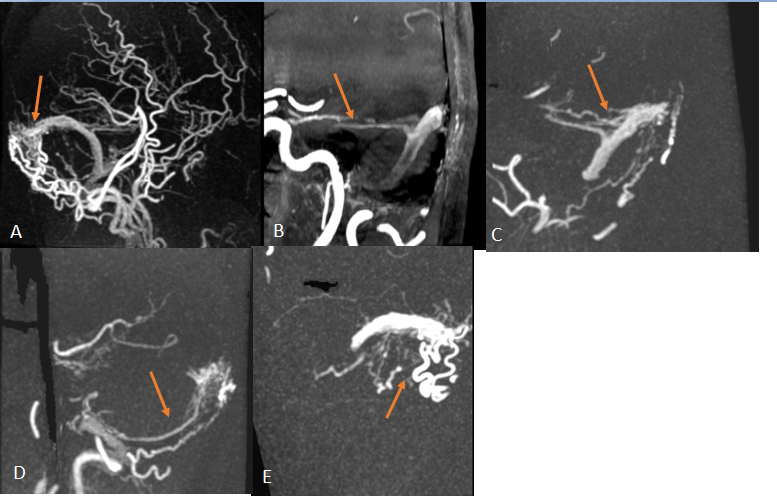
Dural investigated with a new non-contrast MR angiography sequence-silent MRI. The dural fistula is clearly demonstrated with feeders, location and draining vein. Composite angiogram shows dural fistula (arrow in A).The individual arteries can be detected, shown by arrows.(meningohypophyseal artery(B), middle meningeal artery(C), Posterior meningeal artery(D), occipital branches(E)
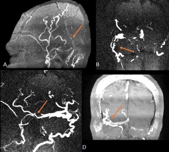
Dural investigated with a time of flight MR sequence. The dural fistula is evident with feeders, location and draining vein, but not as exquisite as silent MRA (A). Images shows occipital artery (B), middle meningeal artery(C), Venous drainage into transverse sinus and cortical reflux can be appreciated(D)
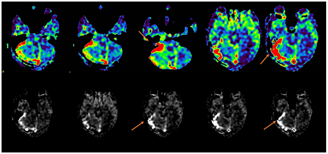
Arterial spin labeling MRI- A new tool for investigating dural fistula. It can detect very small fistula and help in grading of dural fistula.
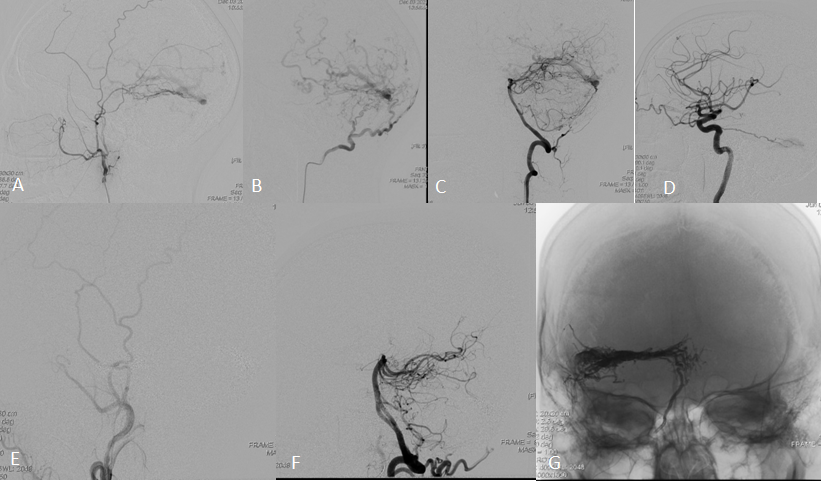
Definitive diagnosis is by angiogram (A-D). It corroborates with previous non-invasive MRI angiogram. The fistula was occluded by endovascular approach (E,F). The embolic cast is evident in G.